MYCOTOXINS IN FEED AND
PIGLETS EAR NECROSIS:
CAUSAL RELATIONSHIP, COFACTORIAL ROLE,
OR COINCIDENCE.
OBJECTIVE OF THE ARTICLE
To critically discuss whether the presence of mycotoxins in piglet feed can be related to ear necrosis, differentiating causal evidence, biological plausibility, and cofactorial role.
EXECUTIVE SUMMARY
The hypothesis that feed mycotoxins contribute to ear necrosis in piglets (Porcine Ear Necrosis, PEN) is reasonable from a biological standpoint, but direct causal evidence is limited. The specific literature on ear necrosis in piglets strongly supports a multifactorial etiology, involving oral behavior, local trauma, lesion microbiota and environmental or management factors [2-6,14].
The first signal that brought mycotoxins into the discussion was the preliminary study by Weissenbacher-Lang et al. [1]. However, later field studies and lesion-characterization studies found low or very low concentrations of mycotoxins in feed or plasma and did not confirm a robust relationship between mycotoxins and PEN severity [3,4].
Among mycotoxins, ergot alkaloids are the exception with the greatest direct plausibility for acral lesions, because their classic mechanism is peripheral vasoconstriction. EFSA considers that the main effects of ergot alkaloids are related to vasoconstriction and hypoprolactinemia, and describes that vasoconstriction may cause gangrenous ergotism with loss of extremities, including ear tips and tail tips [7]. This pathophysiological basis has also been reviewed in depth by Klotz [8].
For DON, fumonisins, aflatoxins, ochratoxin A, T-2/HT-2 and zearalenone, the link with PEN is more indirect: immunomodulation, alteration of the intestinal barrier, changes in microbiota, lower feed intake, poorer tissue recovery, or greater susceptibility to infections [9-11,13]. These mechanisms may turn mycotoxins into cofactors, but they do not demonstrate that they are the primary cause of classic PEN.
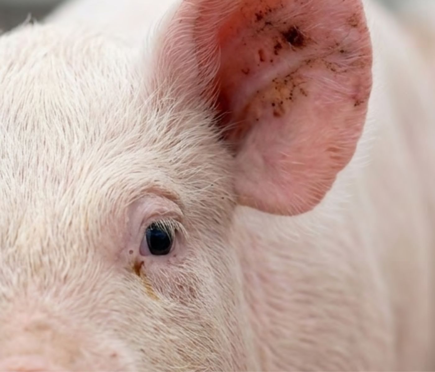
Ear necrosis in post-weaning piglets is observed as dry scabs or ulcerative lesions on the edge of the pinna, which may progress to wet, bleeding lesions and partial tissue loss. Under field conditions, it has been associated with cutaneous and oral bacteria, nibbling, environmental humidity, stress, stocking density, air quality, health status and nutritional factors [2-6,14].
THE PROBLEM
A LESION WITH TOO MANY POSSIBLE EXPLANATIONS
Oral trauma & nibbling
Direct oral interactions represent a critical primary source of ear margin tissue damage, giving opportunistic skin bacteria an immediate pathway to invade.
Feed Mycotoxin
Ergot alkaloids represent a direct vasoconstricting etiology, while DON acts primarily by altering intestinal tissue recovery and dampening physiological immunity.
Lesion Microbiota
Lesions are rapidly superinfected with complex opportunistic bacteria. Inoculation studies show Fusobacterium necrophorum is a primary contributor to advanced necrosis.
Enviroment
Inadequate relative humidity, toxic air contaminants (excess ammonia), lack of space, and thermal stress reduce tissue resistance and increase ear-biting aggression.
Nutrition & Stress
Subclinical intestinal inflammation and post-weaning starvation phases followed by overfeeding alter systemic inflammatory parameters and trigger acral lesion pathways.
Three clinically overlapping conditions should be distinguished. Classic PEN mainly affects piglets after weaning and is usually limited to the ear. Swine inflammation and necrosis syndrome (SINS) is broader, with inflammation or necrosis of the tail, ears, teats, coronary bands, foot pads and other acral areas, even in early stages of life [12]. Gangrenous ergotism, in contrast, is a systemic vascular intoxication that may cause loss of extremities, including the tips of ears and tail [7,8].
This separation is important because the same macroscopic lesion may result from different mechanisms. The presence of mycotoxins in feed is not sufficient to diagnose a mycotoxin-related etiology: it is necessary to demonstrate relevant exposure, epidemiological compatibility, coherent histological lesions, and reasonable exclusion of other etiological pathways [2,4,7].
The document on Verotoxin Associated Syndrome, VAS, [17] and nutritional control provides [19] a complementary hypothesis: some auricular lesions may follow a non-mycotoxic vascular-intestinal pathway, mediated by endothelial damage, increased intestinal permeability, and secondary involvement of Fusobacterium necrophorum, Streptococcus or other agents. This hypothesis does not replace mycotoxin diagnosis, but it prevents the automatic attribution of any acral lesion to feed contamination.
A practical position should be that mycotoxins must be investigated on farms with PEN, especially ergot alkaloids and DON, but diagnosis should be integrated with behavior, histology, bacteriology or metagenomics, assessment of E. coli Stx2e/Vt2e, and a nutritional and environmental audit. [4-7].
2. WHAT THE SPECIFIC LITERATURE ON PEN CONTRIBUTES
The study by Weissenbacher-Lang et al. [1] investigated infectious agents and mycotoxins in piglets with ear necrosis syndrome. It was important because it opened the door to considering DON and ergot alkaloids as possible factors in PEN, but its preliminary design does not allow that signal to be converted into causality.
Subsequent work by Malik and colleagues has shifted the center of gravity toward a multifactorial etiology with a strong behavioral and local component. In prevalence and lesion-characterization studies, mycotoxins measured in feed and plasma were low or very low and were not clearly associated with the presence or severity of PEN [3,4]. Sequencing and lesion studies showed a complex microbiota, with involvement of bacteria such as Fusobacterium, Streptococcus, Staphylococcus and other genera, reinforcing the interpretation of a locally colonized or superinfected lesion [4].
In 2024, Malik et al. showed that oral manipulations of the ear preceded the development of PEN one or two weeks later [5]. Along the same lines, Boulbria et al. in 2024 related PEN prevalence and severity to social behaviors, oral manipulation and markers of inflammation or oxidative stress [6]. These references do not rule out a role for mycotoxins, but they make it unlikely that, in most outbreaks, mycotoxins are the only cause.
The partial reproduction of ear lesions by bacterial inoculation, especially with approaches focused on Fusobacterium necrophorum, supports local infectious involvement and the need for an entry portal or a previously predisposed tissue [15,16]. At this point, both the external hypothesis – trauma, nibbling and infection – and internal or systemic hypotheses that create vascular or intestinal predisposition fit. [17]
Evolution of the evidence
2012 – First Signal: Weissenbacher-Lang et al.
First preliminary study of infectious agents & mycotoxins. Established interest but lacked direct causality proof.
2016 – Cofactor Characterization: Pierron et al.
Described DON & fumonisins as intestinal and immunological cofactors rather than direct primary causes.
2021–2023 – The Paradigm Shift: Malik et al.
Extensive surveys showed weak association of standard mycotoxins with PEN. Complex microbiota highlighted.
2024–2025 – Behavior and Vt2e Confirmation: Malik, Boulbria, & Jordà
Confirmed behavior (nibbling) precedes lesion emergence. EFSA released new strict toxic thresholds for ergot alkaloids.
3. WHICH MYCOTOXINS ARE MOST PLAUSIBLE?
The answer depends on whether the question concerns direct causality or indirect contribution. If direct causality for ear-tip necrosis is required, ergot alkaloids are the strongest candidates because their mechanism is vasoconstrictive and they can produce acral gangrenous lesions [7,8]. If a cofactorial role is accepted, DON, fumonisins, aflatoxins, ochratoxin A, T-2/HT-2 and zearalenone may contribute through less specific pathways related to the intestine, immunity, feed intake and tissue recovery [9-11,13].
Vasoconstriction & ischemic gangrene — VERY HIGH
Intestinal barrier & immunosuppression — HIGH
Intestinal barrier & general health — MEDIUM
Estrogenic transfer / SINS marker — LOW
DON and fumonisins have a plausible pathophysiological connection with the intestinal barrier. The reviews by Pierron, Alassane-Kpembi and Oswald describe how DON and FB1 can alter the intestinal epithelium, modulate the immune response, reduce feed intake and promote greater susceptibility to infections [9,10]. This type of effect may facilitate the progression of lesions initiated by trauma or bacteria, but it is not equivalent to demonstrating PEN caused by DON or fumonisins.
Aflatoxins and ochratoxin A are relevant mainly because of immunotoxicity and systemic effects. Zearalenone is less specific for PEN; its interest is more related to sow-to-piglet transfer, reproductive effects and cases of neonatal acral necrosis or SINS than to classic post-weaning PEN [11-13]. T-2/HT-2 and other trichothecenes have cytotoxic and irritant potential, but the specific evidence for post-weaning ear necrosis is weak.
4. ERGOT ALKALOIDS
THE STRONGEST TOXIC-VASCULAR ARGUMENT
The 2024 EFSA report on ergot alkaloids in feed is especially relevant because it provides an updated toxicological basis. Alkaloids from Claviceps and Epichloë act on vascular and endocrine receptors, with vasoconstriction as a central mechanism. EFSA summarizes that vasoconstriction may lead to gangrenous ergotism, with loss of extremities such as hooves, ear tips and tail tips [7].
This evidence does not mean that every PEN outbreak with mycotoxins is ergotism. PEN affecting post-weaning piglets usually presents epidemiology, prior behavior and lesion microbiota that do not always fit a pure vascular intoxication [2-6]. However, if the feed contains rye, wheat, triticale or cereal by-products at risk of Claviceps contamination, and if there are symmetric or simultaneous acral lesions on tail, ears and limbs, ergot alkaloids must become a diagnostic priority [7,8].
In practice, analysis should include the 14 main alkaloids of C. purpurea and be expressed as a total sum. EFSA considers 0.6 mg/kg of complete feed as a reference point for adverse effects in pigs and piglets [7]. This value is not a “clinical PEN threshold”, but it is a useful reference to decide whether exposure is compatible with a toxic-vascular risk.
RECEPTOR BINDING ALPHA-ADRENERGIC COUPLING
Ergot Alkaloid Ingestion
Alkaloids bind selectively to peripheral adrenergic receptors.
VASOCONSTRICTION ARTERIAL SPASM & LUMEN REDUCTION
Reduced Arterial Flow
Sustained smooth muscle contraction
ACRAL ISCHEMIA DRY GANGRENE & SCAB STAGES
Cell Death & Cyanosis
Loss of tissue elasticity, borders
5. HOW DOES VT2E VEROTOXIN FIT INTO THE PRESENTATION OF PEN?
Studies on the relationship between PEN and VAS and nutritional control provide a complementary idea: not all lesions that appear toxic-vascular should be attributed to mycotoxins [17]. According to these studies, E. coli verotoxin 2e can produce microangiopathy, endothelial damage, increased intestinal permeability and predisposition to acral lesions or to secondary invasion by opportunistic bacteria [17,18].
The study by Jordà et al. in 2025 mentions a reduction in ear necrosis lesions in groups vaccinated against Vt2e and proposes that vaccination would maintain vascular and intestinal integrity [18]. This information should be used as clinical context and as a working hypothesis on farm: if there is evidence of E. coli F18/Stx2e, subclinical edema disease, growth retardation or lack of uniformity, VAS should be included in the differential diagnosis together with mycotoxins [17, 18].
In addition, the study by Ferrando et al. in 2014 highlights carbohydrate availability as a modulator of S. suis virulence [19]. This point is useful for practical management: transition diets that reduce abrupt changes in feed intake, avoid excess poorly digested starch and maintain intestinal stability could reduce the opportunity for bacterial invasion, although this reasoning should be considered complementary and not direct proof of causality [19].
6. DIAGNOSTIC INTERPRETATION ON FARM
In an outbreak, the question should not be “is it mycotoxins or not?”, but rather “what combination of factors has made the lesion possible?”. The first clinical separation should be between a presentation of classic post-weaning PEN, SINS, ergotism and VAS/subclinical edema disease [2,7,12,17, 18].
Histology of recent lesions is decisive to determine whether vasculitis, thrombosis, ischemic necrosis, epidermal trauma or secondary infection predominates [4,14-16].
Feed sampling should be carried out on the batch actually consumed in the two to four weeks before lesion onset, not only on the feed available on the day of the visit. It is advisable to analyze DON, ZEA, fumonisins, T-2/HT-2, ochratoxin A, aflatoxins and a complete panel of ergot alkaloids by LC-MS/MS. When ergotism is suspected, the sum of alkaloids should be compared with risk references for pigs, using EFSA as a framework [7].
Etiological diagnosis of the lesion requires sampling fresh ears, before advanced necrosis or antimicrobial treatments distort the result. The most informative combination is histopathology, targeted bacterial culture, PCR or metagenomics if available, and comparison with unaffected animals from the same batch [4,14-16].
7. PRACTICAL IMPLICATIONS FOR PREVENTION
If analysis confirms ergot alkaloids at relevant levels, the intervention should focus on removing the batch, reviewing high-risk raw materials, cleaning feed circuits and controlling the origin of the cereal. In these cases, general mycotoxin binders should not be considered a sufficient guarantee, because ergot alkaloids do not behave like aflatoxins and their control depends mainly on preventing entry of the contaminant [7,8].
If DON, fumonisins or other mycotoxins predominate at subclinical levels, the strategy is to reduce the total burden, improve digestibility, limit interactions with infections and reinforce intestinal integrity. Specific adsorbents or biotransformers may be meaningful here, but always accompanied by a review of raw materials, particle size, heat treatment, post-weaning feed intake and health status [9,10].
When the evidence suggests that the causes may be related to behavioral modification and the microbiota, the most important measures are to enrich the environment, reduce mixing and weaning stress, ensure adequate feeder and drinker space, control humidity, maintain thermal stability and review the feed-intake curve [5,6].
If there are also indications of Vt2e/VAS, the diagnosis, the history of edema disease and the vaccination program should be evaluated [17, 18].
8. SYNTHESIS OF THE EVIDENCE
Table1. The following tables summarize the relative weight of the main lines of evidence.
The key is not to confuse biological plausibility with causal demonstration.
| Line of evidence | Main finding | Interpretation | Weigth of mycrotoxins |
|---|---|---|---|
| Weistsenbacher-Lang et al. [1] | Preliminary investigation of infectious agents and mycotoxins in PEN. | Generates the mycotoxin hypothesis, especially for DON/ergot, but without demonstrating causality. | Low-moderate. |
| Malik et al. [2-4] | Low mycotoxins in feed/plasma and no clear association with severity. | Weakens the primary-cause hypothesis at low exposures. | Low for direct causality. |
| Malik et al. and Boulbria et al. [5,6] | Oral manipulation and social behavior precede or are associated with PEN. | Strong support for local trauma and bacterial progression. | Indirect |
| EFSA CONTAM and Klotz [7,8] | Ergot: vasoconstriction, hypoprolactinemia and loss of extremities such as ear tips. | Solid toxic-vascular basis for ergotism, not necessarily for classic PEN. | High for ergot as a differential. |
| Pierron et al. [9,10] | DON/FB1 alter intestine and immunity. | Plausible cofactor in the presence of infection or stress. | Moderate as a cofactor. |
| VAS/Vt2e [17, 18] | Non-mycotoxic vascular-intestinal hypothesis with possible bacterial interaction. | Broadens the differential diagnosis and avoids simplistic attribution to mycotoxins. | Complementary |
Table2.The following table differentiates mycotoxins by clinical plausibility in ear necrosis.
| Group | Plausible mechanism | Relationship with PEN | Analytical priority |
|---|---|---|---|
| Ergot alkaloids | Peripheral vasoconstriction; gangrenous ergotism [7,8]. | The most direct relationship with loss of ear/tail tips. | Very high if there are high-risk cereals or acral lesions. |
| DON | Intestinal damage, lower feed intake, immunomodulation [9,10]. | Plausible cofactor; preliminary signal, not confirmed [1,4]. | High |
| Fumonisins | Alteration of intestinal barrier and immunity [9,10]. | Indirect cofactor. | Medium-high. |
| Aflatoxins/OTA | Immunotoxicity and poorer response against infections [10]. | Indirect; no specificity for the ear. | Medium. |
| Zearalenone | Estrogenic effects and sow-to-piglet transfer [11]. | More relevant for reproduction/neonatal disease/SINS than for classic PEN. | Medium if there is compatible clinical evidence. |
| T-2/HT-2 | Cytotoxicity and epithelial irritation. | Plausible as an aggravating factor; specific evidence is weak. | Medium |
9. CONCLUSIONS
The presence of mycotoxins in feed may be related to ear necrosis in piglets as a risk factor or cofactor, but the available evidence does not allow them to be considered a general primary cause of PEN [1-6].
Ergot alkaloids deserve separate treatment: their vasoactive mechanism can produce gangrenous lesions on ear and tail tips, and therefore they should always be analyzed when the epidemiology and raw materials are compatible [7,8].
DON and fumonisins are important because they can alter the intestine and immunity, but their most likely role is to facilitate the progression of lesions induced by behavior, bacteria or other factors, not to initiate typical ear necrosis on their own [9,10].
Studies on VAS/Vt2e reinforce that ear necrosis may have a non-mycotoxic vascular-intestinal pathway. This makes it necessary to include E. coli Stx2e/Vt2e and subclinical edema disease in the differential diagnosis [17, 18].
The best on-farm approach is multifactorial: analysis of mycotoxins and ergot, early histology, microbiology of fresh lesions, Vt2e assessment, nutritional review, and behavioral and environmental audit [4-7,17, 18].
MINIMUM RECOMMENDED PROTOCOL IN A PEN OUTBREAK
1. Characterize the lesion
Characterize the lesion: age at onset, percentage of affected pens, symmetry, presence of tail/hoof/teat lesions, dry or wet evolution, and temporal relationship with feed changes [2,12].
2. Sample the feed consumed before the outbreak
Sample the feed consumed before the outbreak: keep subsamples from the silo, feeder or bag; analyze by LC-MS/MS DON, ZEA, FB1+FB2, T-2/HT-2, OTA, aflatoxins and ergot alkaloids [7,9,10].
3. Take biopsies or pinnae from early lesions
Take biopsies or pinnae from early lesions: histopathology, bacterial culture and, if possible, metagenomics or a molecular panel [4,14-16].
4. Evaluate VAS/Vt2e
Evaluate VAS/Vt2e: E. coli F18/Stx2e, signs of subclinical edema disease, growth retardation, uniformity and response to vaccination if there is a history [17, 18].
5. Audit management
Audit management: stocking density, mixing, enrichment, ventilation, humidity, temperature, feeder/drinker space and fasting/binge-feeding episodes after weaning [5,6,17].
6. Interpret with controls
Interpret with controls: compare affected and unaffected animals from the same batch and use data from previous batches to avoid erroneous attribution to a single factor [3,4].
The central message of the article is that piglet ear necrosis should be approached as a multifactorial condition. Mycotoxins may contribute to the process, but exposure, lesion characteristics, microbiology, behavior, environment, nutrition and alternative vascular mechanisms must all be evaluated together.
🔒 Contenido exclusivo para usuarios registrados.
Regístrate gratis para acceder a este post y a muchos más contenidos especializados. Solo te llevará un minuto y tendrás acceso inmediato.
Iniciar sesiónRegístrate en nutriNews
REGISTRARME